
The main joint of the shoulder is called the glenohumeral joint. Broadly speaking, its main components are the head of the humerus (the upper end of the arm bone) and the scapula. This join plays an essential role in the mobility of the forearm and its shape can provide information about the locomotion of a given species. In this context, is particularly interesting the study of the structure that articulates with the head of the humerus, called glenoid fossa, or simply, glenoid. From the size and the shape of the glenoid it can be inferred the postural and locomotor activity of a species, extant or extinct.
Although the relationship between the shoulder bone morphology and its function is well known, very little is known about the involvement of the soft tissue (such as cartilage, tendons, or connective tissue). In this context, there has been debate within biological anthropology about what role the cartilage in general, and the cartilage that surrounds the glenoid cavity in particular, can play in the locomotor behavior of primates. This cartilage is called the glenoid labrum or simply labrum and it is an essential part of the glenohumeral joint. Despite its scientific interest, few morphological studies have been carried out on the glenoid labrum in primates and humans due to the difficulty in collecting quantitative information of cartilaginous structures, since soft tissue is not preserved in fossils and in extant species due to the difficulty of accessing cadaveric samples.
Now, a recently published paper in the American Journal of Biological Anthropology led by Georgina Raventós-Izard (predoctoral researcher, Paleoprimatology and Paleoanthropology, ICP; PhD student, Geology program, UAB) has carried out a pioneering study of the morphology of the labrum using a leading technique, the photogrammetry. The aim of the study was to determine the effect of the labrum on the locomotor and postural behavior of extant primates and humans to infer it in fossil primates and hominins.
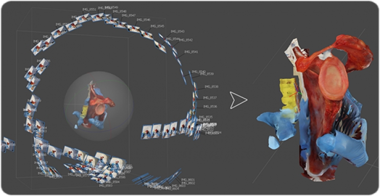
Photogrammetry is a technique that consists of the three-dimensional (3D) reconstruction of different structures from photographs taken from different angles. The reconstruction software then uses the common points of the images to reconstruct a 3D model of the structure. Thanks to this technique, it was possible to create 3D virtual models of glenoid cavities with the labrum of different individuals. Later, thanks to geometric morphometry techniques, the shape of the models was quantified, and their morphological variation was visualized.
The study concludes that making inferences about the locomotor behavior of primates based on the morphology of the glenoid cavity without taking the cartilage into account could let to errors. In other words, the morphology of the glenoid cavity does not determine the functionality on its own and the labrum should be of the considered in this type of studies.
Main image: Process of reconstructing a 3D model, using photogrammetry, of a human scapula displaying the glenoid cavity with the presence of the glenoid labrum.
Original article:
- Raventós-Izard, G., Potau, J. M., Casado, A., Pastor, J. F., & Arias-Martorell, J. (2023). The morphofunctional implications of glenoid labrum of the glenohumeral joint in hominoids. American Journal of Biological Anthropology, 181, 195-205. https://doi.org/10.1002/ajpa.24729


